
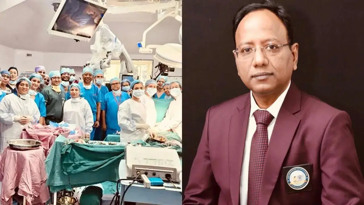
New Delhi: In the realm of medical science, when determination, modern technology, and the extraordinary expertise of doctors converge, even the seemingly impossible can become reality. Doctors at the All India Institute of Medical Sciences (AIIMS), New Delhi, have achieved such a historic feat. They have written a new chapter in India’s medical history by performing a complex, in-utero (inside the womb) surgery to correct a severe congenital spinal deformity in a fetus that had not yet been born.
On November 12, AIIMS successfully performed its first open fetal surgery to treat myelomeningocele—the most severe form of spina bifida—in a fetus at the 25th week of gestation. During this highly challenging six-hour procedure, the exposed portion of the fetus’s spine was repaired while still inside the womb. In this condition, the spinal cord and its protective membranes protrude outside the body, potentially leading to the leakage of cerebrospinal fluid. Without timely intervention, the child could face paralysis, weakness in the legs, hydrocephalus (accumulation of fluid in the brain), and lifelong, severe physical complications.
Five Weeks of Preparation, A Decisive Six-Hour Operation
This was no ordinary surgery. It was the culmination of over five weeks of intensive preparation, involving multiple rounds of expert consultations, detailed fetal examinations, the use of advanced imaging technology, counselling for the mother and her family, specialised anaesthesia protocols, and comprehensive rehearsals of the procedure.
The neurosurgical component of the operation was led by Professor Deepak Gupta from the Department of Neurosurgery at AIIMS. The Department of Maternal-Fetal Medicine and Obstetrics was overseen by Professor Neena Malhotra and Professor K. …was handled by Aparna Sharma. The obstetric and neuro-anesthesia team during the operation included Professor Vimi Rewari, Professor Mihir Pandia, Dr. Rashmi Ramachandran, and Dr. Gyanendra Pal Singh.
Dr. Shashank Chauhan from the Department of Plastic Surgery played a key role, while the neonatology team included Dr. Ramesh Agarwal and Dr. Deepika Kanth.
The patient’s initial evaluation and counseling were conducted by Professor K. Aparna Sharma, along with Dr. Anubhuti Rana, Dr. Deepali Garg, and Dr. Saloni Kamboj. Dr. Smita Manchanda from the Department of Radiodiagnosis and Interventional Radiology provided significant support in developing a detailed pre-operative plan.
Surgery under the guidance of high-tech microscopes and ultrasound
This complex procedure was performed at the AIIMS Neurosciences Centre, utilizing state-of-the-art microscopes and continuous ultrasound monitoring. Throughout the operation, the condition of the fetus was closely observed to ensure the safety of both mother and child. More than 30 doctors and healthcare professionals—from the departments of Obstetrics and Gynecology, Maternal-Fetal Medicine, Neurosurgery, Obstetric Anesthesia, Neuro-anesthesia, Plastic Surgery, Radiodiagnosis, Neonatology, and Nursing Services—participated in this landmark undertaking.
What is Myelomeningocele?
Myelomeningocele is the most severe form of spina bifida. It is a congenital defect in which the lower part of the fetal spinal cord fails to close completely. As a result, the spinal cord and its protective membranes protrude outward. This condition carries a risk of damage to the nervous system. After birth, such children may face complications including weakness or paralysis in the legs, issues with bladder and bowel control, the accumulation of fluid in the brain (hydrocephalus), and other neurological challenges.
In India, the burden of neural tube defects—congenital malformations related to the neural tube—is significant. A systematic review of 27 Indian studies, published in 2024, estimated the combined prevalence of these defects at 9.46 per 1,000 births. However, rates vary across different regions, highlighting the need for a more robust surveillance system in the country.
Experts clarify: It is a form of treatment, not a miraculous, complete cure.
Professor Deepak Gupta from the Department of Neurosurgery at AIIMS—who also serves as the President of the Indian Society for Pediatric Neurosurgery—described this achievement as the emergence of new capabilities in the fields of fetal medicine and pediatric neurosurgery in India. According to him, the objective of performing surgery while the fetus is still in the womb—in select cases—is to protect exposed neural tissue from further damage, reduce the likelihood of needing a shunt (to drain fluid from the brain) after birth, and improve certain neurological outcomes. It is, however, not a complete cure for the condition. These children may continue to require long-term specialised care and multidisciplinary medical support. Professor Neena Malhotra noted that this achievement is the result of the collective dedication of a multidisciplinary team possessing exceptional expertise.
Meanwhile, Professor Aparna Sharma explained that the decision to perform this operation was made with great caution and detailed planning, keeping in mind the best possible health outcomes for both the mother and the child. She noted that such surgeries require extensive counseling, high-level anesthesia, and continuous monitoring of both the mother and the fetus—both before and after the procedure.
International research also validates the importance of in-utero surgery
International studies support the significance of performing surgery for myelomeningocele while the fetus is still in the womb. The ‘Management of Myelomeningocele Study’ (MOMS) conducted in the United States revealed that among children who underwent in-utero surgery, only 40 percent required a shunt (to drain cerebrospinal fluid) by the age of 12 months, compared to 82 percent among those who underwent surgery after birth. The study also observed improvements in certain motor functions and a reduction in the hindbrain malformation associated with Chiari II malformation in children who received in-utero surgery.
However, this operation carries risks, including the possibility of preterm labor, complications related to surgical scarring on the mother’s uterus, and potential health risks to the fetus. Therefore, this procedure should only be performed in carefully selected cases and at specialized centers equipped with state-of-the-art facilities.
No financial burden on the family
A significant aspect of this landmark operation was that the family did not have to bear any costs. The procedure was provided free of charge under the Government of India’s ‘Janani Shishu Suraksha Karyakram’ (JSSK). According to AIIMS, a comprehensive medical package—encompassing fetal surgery, high-risk obstetric care, delivery, and neonatal treatment—could cost over 10 million rupees (1 crore INR) in the United States, depending on the specific medical requirements of the patient.
Doctors’ appeal: Folic acid is essential for preventing the condition
Experts at AIIMS clarified that while in-utero surgery is a vital treatment option, it cannot serve as a substitute for the prevention of the condition. The formation and closure of a baby’s neural tube are completed within approximately 28 days of conception—a process that often occurs before a woman even realizes she is pregnant. Therefore, it is crucial to seek medical advice regarding folic acid intake prior to conception, ensure an adequate supply of folate in one’s diet, and implement the scientific fortification of food products with folate. Furthermore, there is a need to develop a robust, nationwide surveillance system for such congenital anomalies.
A dream envisioned 14 years ago has now become a reality
This achievement by AIIMS is not limited to a single successful operation; it opens doors to new possibilities for modern fetal medicine in India. According to the institute’s team, both the mother and the fetus will remain under the joint supervision of specialists for the remainder of the pregnancy, during delivery, and even after the baby is born. With this milestone, AIIMS New Delhi has demonstrated that, for carefully selected families, the option of undergoing surgery for myelomeningocele while the baby is still in the womb is now available in India.
Medical experts note that the possibility of performing fetal spinal surgery was first discussed at AIIMS about 14 years ago. What was once a dream has now become a reality. The primary challenge ahead lies in ensuring that this technique remains safe, effective, and accessible to Indian families in need. In this procedure, even before the child has entered the world, doctors have already begun the fight for their better future. Truly, this is a triumph for medical science—a battle for life fought even before birth.